Companion Animal

See the Unseen

Equine Thermography

Equine patients cannot tell us where they have an issue.  Digatherm’s thermal imaging system solves that problem by identifying areas that would benefit from further examination. These images provide real-time information about the circulatory, musculoskeletal, and nervous systems.

what-is-equine-thermal-imaging
what-is-equine-thermal-imaging-2

What is Equine Thermal Imaging?

Equine Thermography – a  technique to evaluate patients

Equine thermal imaging uses a medical-grade infrared camera to measure the temperatures emitted from the body surface.  The software converts temperature data into images that evaluate the patient’s physiology.  These color images provide a physiological map of the patient. This allows veterinarians to see thermal patterns and impressions unique to that patient.  Any abnormal pattern warrants further evaluation.

The Digatherm® medical-grade infrared imaging system equipped with veterinary-specific software makes reading thermal images intuitive for clinicians.  With colors representing temperatures in the thermal image, clinicians can easily identify unexpected areas of increased or decreased temperatures.  Each of these areas warrants further investigation and consideration of etiology.


spec

Medical-grade accuracy

spec

The pioneer in veterinary thermography

spec

Appropriate for every type of veterinary practice

spec

Comprehensive and individualized training


How does infrared thermal imaging benefit the equine practice?

Non-invasive medical thermal imaging provides a physiological study of the entire patient. Anatomical areas that would benefit from further evaluation are easily identified. This is possible often before any clinical signs or structural changes exist. In addition to the primary areas of concern, the discovery of all secondary and compensatory areas are identified. Beneficial treatments can simultaneously be administered to all affected areas. This allows the equine patient a shorter recovery time and even the prevention of an injury.

A Digatherm® equine thermal imaging camera objectively monitors any pharmaceutical or modality-based therapeutic plan. The common question: “When can they return to work?” is answered by visualizing the progress of the case.  Early return to work often results in reinjury. Monitoring with thermal imaging helps validate complete healing before return to work.

When clients see thermal images, they better understand their horse.
This understanding supports diagnostic and treatment recommendations. When combined with ultrasonography, radiography, or nuclear scintigraphy, thermal images shorten the path to definitive diagnoses.

  • Detecting issues early before any clinical signs
  • Objective monitoring of any therapeutic or rehabilitation plan
  • Client education allowing better understanding and compliance
Equine-use-case-2

testimonial-quotation-icon

“This must be what it felt like to discover penicillin!  I’ve actually made it home before dark two days in a row, because this system takes me straight to the area of lameness.”

– BRIAN GARRETT DVM, CVSMT


What can you see?

Below are several case examples using equine thermal imaging.

Case 1: Equine Suspensory Ligament Injury

Presentation-1-01-1Presentation-1-03Presentation-1-02


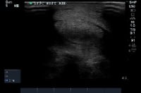
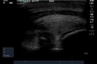
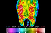
Presentation: Thoroughbred, 2-year-old. Lameness examination.

Digital Thermal Imaging: A physiological exam was conducted capturing images using a Digatherm thermal imaging system.

Interpretation: Areas of hyperthermic activity medially along the entire length of the left metacarpal with a focal area medially at level 3, a focal area proximal to left medial sesamoid, and another focal area over the left lateral sesamoid. The areas of hyperthermia correlate with the lesions on the ultrasound.

Case 2: Kissing Spines

Presentation-1-04-1

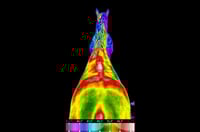
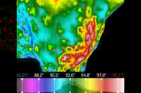
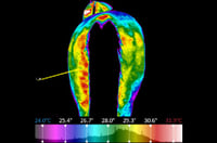
Presentation: Equine patient with back pain

Digital Thermal Imaging: Asymmetrical hyperthermia thoracic and lumbar spine and surrounding musculature

Interpretation: Radiographs show remodeling of thoracic articular processes of T14-16.

Diagnosis: Baastrup’s disease (Kissing Spines)

Case 3: Fracture of the Supraglenoid Tubercle of the Scapula

Presentation-1-05Presentation-1-06Presentation-1-07


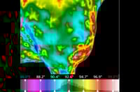
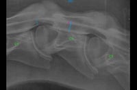
Presentation: Equine patient with undiagnosed lameness. Several lameness exams and radiographic studies failed to localize the issue.

Digital Thermal Imaging: A physiological exam was
conducted capturing anterior to posterior and lateral images of the shoulders.

Interpretation: Hyperthermia repeatable in all views over scapulohumeral joint.

Diagnosis: Repeat radiographs taken of this area revealed a fracture of the supraglenoid tubercle of the scapula.

Credit: Dr. Liz Steele, Steele Equine (Zolfo Springs, FL)

Case 4: Detection of Osteoarthritis of Tarsocrual Joint

Presentation-1-08-1Presentation-1-09Presentation-1-10


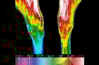
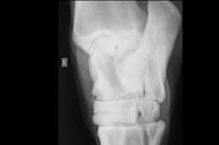
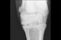
Presentation: Three-year-old Quarter Horse

Digital Thermal Imaging: A physiological exam was
conducted capturing an anterior to posterior image of the hocks.

Interpretation: Bilateral generalized hyperthermia over proximal aspect of hocks, with focal assymetry on the anterior medial aspect of the left hock (location 1).

Diagnosis: Radiographs confirm left tarsal osteoarthritis at the location of most intense hyperthermia.

Case 5: Focal Hypothermia in a Healing Wound

Presentation-1-12Presentation-1-11


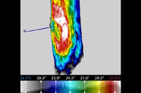
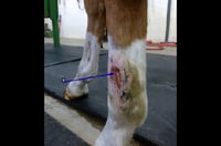
Presentation: Five-year-old American Quarter Horse with a traumatic wound that dehisced 21 days after primary closure.

Digital Thermal Imaging: The non-healing wound was imaged at day 37.

Interpretation: The thermal image showed asymmetry, with expected hyperthermia proximal, distal and posterior to the wound, but an abnormal area of hypothermia was observed on the anterior aspect of the wound.

Diagnosis: Differential diagnoses included sequestrum, foreign body, or other cause of delayed healing.

Credit: Chelsie McAllister, DVM, Valley Veterinary Clinic (Glasgow, MT)

Case 6: Cervical Vertebral Stenotic Myopathy

Presentation-1-21Presentation-1-17Presentation-1-34


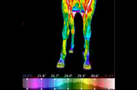
Presentation: Yearling Thoroughbred with acute lameness and mild ataxia

Digital Thermal Imaging: A series of screening images were taken of the entire colt to look for thermal asymmetry.

Interpretation: Focal hypothermia in the distal lateral left forelimb indicated a need for neurological assessment of the cervical region. Hyperthermia over the caudal aspect of the left hindlimb suggest possible inflammatory response typical with compensatory process.

Diagnosis: Radiographs detected cervical vertebral stenosis C3-C4.

Digatherm® Thermal Imaging Systems

Learn more about Digatherm by ICI's veterinary tools and technology.

IR Pad 640

Our award-winning IR Pad 640 provides medical-grade resolution, accuracy, and sensitivity to the veterinary market. Veterinary specific software and calibration provide high-quality thermal images that are easily interpreted.

IR Pad 320

Our IR Pad 320 is easy to use in any veterinary practice. The handheld tablet allows vets to quickly visualize and document changes in a patient's physiological state in real-time.

Request a demonstration.

Discover the Digatherm by ICI difference and learn more about the most innovative and accurate thermal imagers available. Fill out the form below to request a demonstration of our state-of-the-art thermal camera.